Unraveling Molecular Secrets
Scientists around the globe use microscopy technique invented by UC Irvine researcher
- Photo-induced force microscopy, a technique that enables imaging of specimens at the nanometer scale, was developed by a UC Irvine researcher nearly 20 years ago.
- Since its invention, PiFM has become a research tool of choice for biologists, materials scientists, geologists and others in nearly every corner of the world.

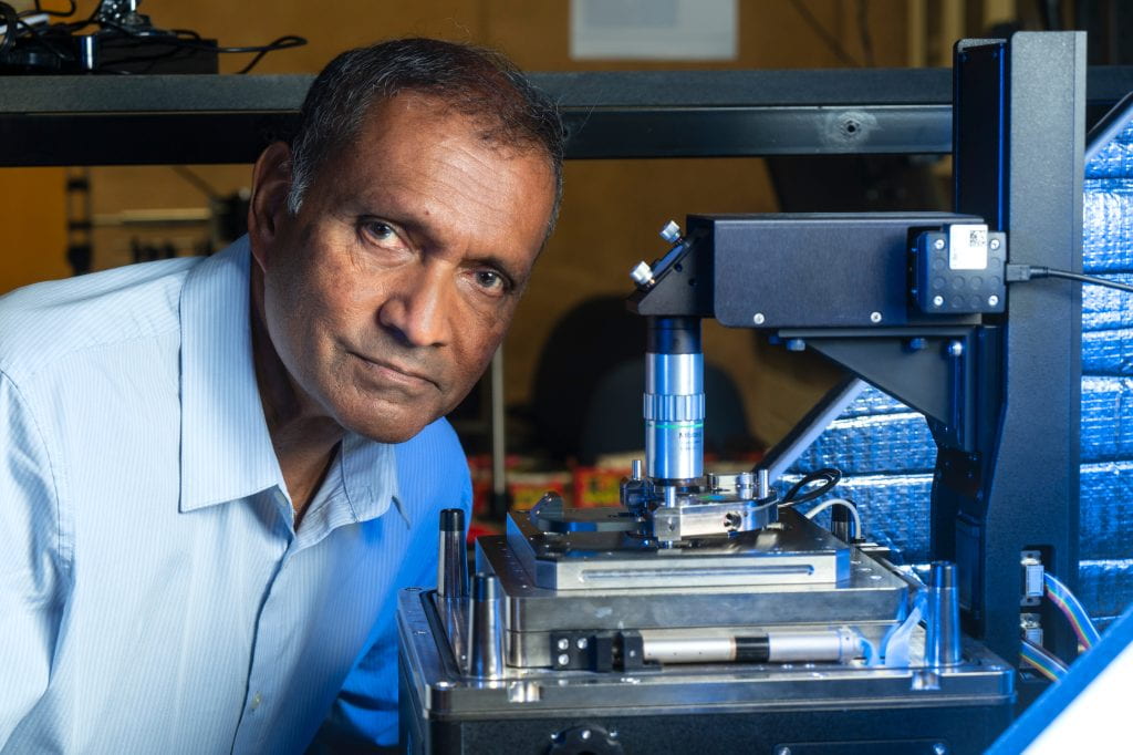
July 11, 2025 - Photo-induced force microscopy began as a concept in the mind of Kumar Wickramasinghe when he was employed by IBM in the early years of the new millennium. After he came to the University of California, Irvine in 2006, the concept evolved into an invention that would revolutionize research by enabling scientists to study the fundamental characteristics of matter at nanoscale resolution.
Since the earliest experimental uses of PiFM around 2010, the device, which reveals the chemical composition and spatial organization of materials at the molecular level, has become a tool of choice for researchers in fields as diverse as biology, geology, materials science and even advanced electronics manufacturing.
“This is the story of a technology that was inspired by work at IBM, was invented and developed at UC Irvine, then got spun off, and now we have instruments on all continents across the world except for Antarctica,” says Wickramasinghe, Henry Samueli Endowed Chair and Distinguished Professor emeritus of electrical engineering and computer science who now holds the title of UC Irvine Distinguished Research Professor. “Almost anywhere serious research is happening, there are people out there who are using PiFM to discover new things.”
To commemorate the invention of PiFM and its proliferation in the scientific community, Nature Reviews Methods Primers recently published an article outlining the technology’s capabilities and applications, with Wickramasinghe and an international team of colleagues as co-authors.
Wickramasinghe says the initial work on PiFM at UC Irvine was made possible through support he received from the Samueli Foundation and unrestricted funding from the W.M. Keck Foundation.
“That’s really helpful because when you have crazy ideas like this, it’s very hard to get the National Science Foundation or some other agency to support it, because they want to see results first before they give you money,” he says.
Eventually, however, the now-discontinued NSF-funded Center for Chemistry at the Space-Time Limit at UC Irvine did provide backing. Its researchers shared the vision that by combining nonlinear optics with scan-probe microscopy, it should be possible to visualize molecules in action, in atomistic detail.
“Atomic force microscopy, which Professor Wickramasinghe had pioneered, was central to the research we pursued in CaSTL,” says V. Ara Apkarian, the center’s director. “Upon learning about the PiFM work Kumar was carrying out across the campus, we immediately invited him to join the center and initiated collaborative efforts that would last until the sunset of the center in 2019. It was serendipity, but we were fortunate to have in our midst Professor Wickramasinghe, whose newly invented PiFM had tremendous promise toward our goals.”
Apkarian, UC Irvine Distinguished Professor emeritus of chemistry, says that the first space-time-resolved PiFM images on the nanometer-picosecond scale were the result of a multi-investigator effort that appeared in print in 2015.
From lecture notes to a usable instrument
Wickramasinghe says the inspiration for the photo-induced force microscopy technology originated in notes he prepared for a lecture on semiconductor physics. He says that metal-semiconductor junctions found in practically all chips have the potential to become diodes like the ones used in solar cells.
According to Wickramasinghe, when a negative voltage is applied to the metal side of an n-type semiconductor-metal junction, almost no current flows, as electrons must overcome a large potential barrier to get to the semiconductor. The current is very small when compared to the electrons moving in the opposite direction.
“When the same negative voltage is applied to the semiconductor side, electrons ‘see’ a much smaller barrier at the junction, resulting in currents orders of magnitude higher,” he says. “In the latter case, there’s an additional effect that further increases the current. As an electron approaches the semiconductor-metal junction, it ‘sees’ its positive charge image on the metal side, and the attractive force between the negative electron and its positive charge image further lowers the barrier for the electron to go across – an effect easily detected in the current.”
Wickramasinghe says he thought that since this electron barrier crossing is readily perceivable, he might be able to directly observe and measure the electromagnetic forces involved in the interaction between an optically driven molecule and its mirror image, which he calls the “image force.”
“A molecule that’s driven externally with light would not be just one charge but a dipole – an oscillating minus and plus charge pair,” he says. “There would be a mirror image on the other side of the junction, a plus/minus charge, as the molecule approaches a metal surface, resulting in an attractive force. We set about trying to detect that force, and our success led to the creation of this instrument.”
How photo-induced force microscopy works
According to Wickramasinghe, there are multiple methods that combine optics and scanning-probe microscopy, classed as “near-field scanning optical microscopy.” These techniques employ a tip or sharp stylus to focus light at the junction of itself and a substrate under interrogation and a detector to measure the intensity of the scattered light. Image contrast is obtained because matter is colored – different molecules respond in unique ways to varying colors of light. Distinct from all other methods, PiFM detects the scattered photons at the junction by the electromagnetic force they exert on the tip. Using clever modulation schemes, the momentum of individual photons is detectable, Wickramasinghe says.
Laura Otter, an Earth sciences research fellow at the Australian National University, says that she uses PiFM in her role as a scientist specializing in biomineralization, the process by which living organisms form minerals. Otter says she’s interested in investigating – at the microscale and the nanoscale – how mollusk shells, otoliths (vertebrate ear bones) and corals grow and respond to changing environments.
“PiFM enables me to zoom deep into my samples and map where organic molecules and minerals meet at the nanometer scale, which is something not possible with other techniques. Using PiFM, I was able to visualize how the mineral component in mother-of-pearl transforms from an amorphous to a crystalline state and how the initial amorphous phase incorporates higher amounts of trace elements than we’d expected,” Otter says. “This finding has important implications for reconstructing past environmental conditions from shell materials.”
In addition to PiFM being used by scientists around the world, there are several such devices on the UC Irvine campus, including in biology, materials science and chemistry labs.
Wickramasinghe says that the capabilities of PiFM go beyond fundamental sciences into the field of advanced electronics and other technologies.
“Because it has spectroscopy capability on the nanoscale, PiFM lends itself to many industrial applications. For instance, you can use it to map and study the chemistry of the most advanced lithographically printed circuits before they’re produced on a mass scale, allowing you to check whether your process is on the right track,” he says. “If you’re using an Apple iPhone, for example, there’s a good chance that the lens on its camera was scanned at some point using the instrument we created here at UC Irvine.”
Wickramasinghe notes: “Before photo-induced force microscopy was developed, there was certainly a gap in science, an inability to conduct infrared spectroscopy on the nanometer scale. It was a hole that I tried to fill for many years. It was a slow process, but it’s gratifying to see how PiFM has caught on and is now aiding in scientific research nearly everywhere.”
- Brian Bell
